General Urology
Exceptional Service & Treatment for General Urologic Conditions
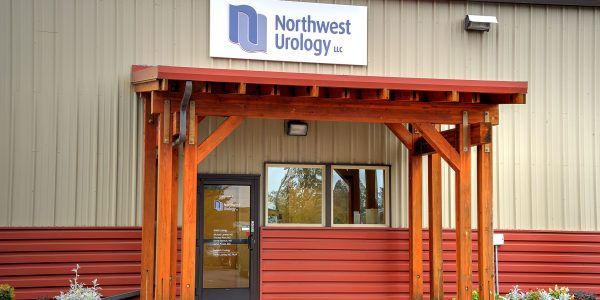
Northwest Urology is a leader in state-of-the-art treatments for all urologic cancers and general urologic conditions. We have expertise in treating the full range of female uro-gynecological conditions, pediatric urology, sexual dysfunction and urologic trauma.
Northwest Urology, LLC provides expert care for the entire spectrum of general urologic conditions. These include:
Stone Disease
The kidneys produce urine as they process waste and extra water out of the blood and maintain the body’s balance of water, salts, acid, and minerals. These substances normally dissolve in the urine, but may become concentrated and form crystals (stones) in the wall of the kidney. There are several different types and sizes of kidney stones.
Kidney stones are most commonly diagnosed when they break away from the wall of the kidney, travel with the flow of urine, and lodge in the ureter. The stone(s) obstruct or restrict the flow of urine and increase pressure in the ureter and kidney. The resulting pain, which is typically felt in the back and lower abdomen/groin, is often excruciating and prompts the patient to seek emergency treatment. Kidney stones can cause other symptoms that include blood in the urine, nausea/vomiting, persistent urinary tract infections and urinary frequency.
Our urologist will perform a thorough evaluation of all symptoms, as well as diagnostic tests to confirm the presence and location of the stone(s). Urine will be tested for infection or evidence of blood, and a blood test will determine the baseline kidney function. X-rays or a CT scan may be performed to provide definitive proof of the stone(s).
Based on the results of the diagnostic tests and examination, we will develop an appropriate treatment plan. Kidney stones that are too large to pass on their own, cause pain that cannot be controlled with oral medication, or are associated with infection, fever or vomiting require urgent treatment. Additionally, patients with poor kidney function, a single kidney or a complete blockage of urine need emergency treatment.
There are a number of treatments available for kidney stones, and we will counsel patients and select the method best suited for their individual condition. The most common treatment options are:
ESWL (extracorporeal shock wave lithotripsy):
- A series of shock waves are used to break the stone into fragments small enough to pass out of the kidney
- Requires no incision and is usually performed as an outpatient procedure under general anesthesia
Ureteroscopy:
- A small telescope is inserted through the bladder and up the ureter to reach the kidney stone
- The stone is fractured using laser energy delivered via a small glass fiber. Fragments of stone are removed by grasping with a stone “basket”
- Typically an outpatient procedure that allows patients to return home the same day; although some patients may need 24-48 hours of observation after the procedure
Percutaneous Nephrostolithotomy (PCNL or “PERC”):
- Used for removal of larger stones that cannot be treated by other means
- A hollow tube is placed into the kidney through a small (1 inch) incision in the back
- An ultrasonic lithotripter (or laser energy) is used to pulverize the stone(s) into granules that can be suctioned from the kidney or removed with graspers
Open Surgery:
- For kidney or ureteral stones – is rarely required
Combination Treatment:
- May be used in patients with especially large stones that are not successfully removed by any one technique
After their removal, the kidney stones are analyzed to determine their chemical characteristics. This information is important for effective post-treatment management, as there are several ways to help prevent kidney stones from recurring. For patients with recurrent kidney stones, analyzing the urine’s molecular components with a 24 hour urine collection will further guide prevention efforts.
Voiding Dysfunction/Urinary Incontinence
The bladder is a hollow muscular organ that stores urine. As it fills, the muscles of the bladder relax, allowing it to expand. Urination is controlled by the sphincter, a muscle at the bottom of the bladder. When the bladder gets full and the situation is appropriate to urinate, the sphincter relaxes and the bladder contracts to allow urine to pass through the urethra without difficulty.
Voiding dysfunction describes abnormal bladder and/or urethral functions. The condition can affect both men and women. It includes the abnormalities associated with the bladder’s ability to store urine or empty properly. Urologists treat the entire range of voiding dysfunctions including incontinence (involuntary release of urine), overactive bladder, and urinary retention.
Overactive Bladder
Overactive bladder is a common problem that causes a sudden urge to urinate. It sometimes leads to Urgency Urinary Incontinence (UUI). Patients describe UUI as the inability to postpone urination even when the bladder is nearly empty. The condition can affect both men and women. One of every six Americans suffers from overactive bladder symptoms, which may also include urinary frequency and nighttime voiding frequency.
The patient’s description of symptoms, complete medical history and physical examination are the initial steps in diagnosing overactive bladder. Urodynamic testing, which provides objective and quantitative measures of bladder capacity and bladder pressures, may be utilized to assist in the diagnosis. (Urodynamics means the study of the transport, storage, and expulsion of urine.)
Video urodynamics may be used to provide a more detailed understanding of the patient’s bladder physiology. Video urodynamics combines traditional urodynamic testing with fluoroscopy (moving X-rays) to gather anatomical information in real-time with subjective input from the patient. Having both functional and anatomic information allows our urologist to develop more individualized treatment strategies.
Effective treatment options for overactive bladder range from prescription medicine to physical therapy to surgical intervention. Many patients will benefit from a conservative (non-surgical) treatment plan.
These treatments include:
- Pelvic floor rehabilitation – The patient performs simple exercises that strengthen the pelvic muscles, also known as Kegel exercises.
- Behavioral therapy – Methods such as bladder retraining or timed voiding can help control urgency.
- Medical therapy
When non-operative intervention is ineffective, the following minimally invasive surgical options may be considered:
- Bladder Botox Injection – Medication is injected into the bladder to help the muscle relax and reduce urgency, frequency and incontinence
Incontinence
Urinary Incontinence (UI) is the accidental release or leakage of urine. Incontinence affects over 14 million Americans and can markedly affect the patient’s quality of life. UI is almost twice as prevalent in women as it is in men.
There are several types of UI with the most common being Stress Urinary Incontinence (SUI), followed by Mixed Urinary Incontinence (MUI) and Urgency Urinary Incontinence (UUI). SUI is the involuntary release of urine due to the contractions of the abdominal muscles when sneezing, laughing, coughing, etc. Patients describe UUI as the inability to postpone urination. MUI is the combination of both SUI and UUI. SUI is rare in men and usually a consequence of prostate cancer surgery.
The patient’s description of symptoms and their impact on quality of life followed by a complete physical examination are the first steps in diagnosing voiding dysfunction. Patients may also complete a questionnaire to help identify the type and severity of incontinence. At the initial consultation, urine analysis and bladder ultrasound are usually performed.
Treatment options for urinary incontinence include:
- Pelvic Floor Rehabilitation – Exercises that strengthen the pelvic muscles, also known as Kegel exercises
- Behavioral Therapy – Methods such as bladder re-training, timed void and double voiding
- Medical Therapy – Medications are available that reduce symptoms of UUI
For more complex cases that fail to respond to the previous therapies, UC Health Urology provides minimally invasive treatments as well as complex surgical procedures for the entire range of incontinence conditions.
Minimally Invasive Procedures include:
- Bladder Botox Injection – medication injected into the bladder to help relax the bladder muscle and reduce urgency
- Sub-urethral Sling – a procedure that restores the urethra to its normal position
- Artificial Urinary Sphincter – an implantable device that replaces the function of the patient’s bladder sphincter
Complex Surgical Procedures include:
- Bladder Augmentation – a procedure that uses a piece of tissue from the bowel, stomach or ureter (the tube that carries urine from the kidney to the bladder) to enlarge the bladder capacity
- Urinary Diversion – diverting the urine flow from the natural urethra to an abdominal stoma (opening)
Neurogenic Bladder
Neurogenic bladder is a condition in which a person lacks bladder control due to a brain or nerve condition such as Alzheimer’s disease, Parkinson’s disease, multiple sclerosis, or stroke. Symptoms include overactive bladder (frequent urination, loss of bladder control) or an underactive bladder (bladder becomes too full due to urinary retention, trouble starting to urinate, or unable to tell when the bladder is full). This condition may be further complicated by recurrent urinary tract infections (UTIs), urinary stones or cancer.
Neurogenic bladder involves the nervous system and the bladder. Our urologist will recommend a variety of tests to determine the status of both. Video urodynamic studies measure bladder capacity, bladder pressure, urine flow, and bladder emptying. Kidney function will be assessed to see if the kidneys have become compromised due to high bladder pressure. Imaging studies of the skull, spine, and urinary tract with x-rays, magnetic resonance imaging (MRI), or computed tomography (CT) complete the evaluation. If necessary, a neurologic evaluation with EEG may be recommended.
The most effective treatment plan is developed based upon a thorough evaluation of all of the test results, voiding symptoms and type of bladder dysfunction. Therapies for neurogenic bladder fall into four categories: behavioral, electrical stimulation, drug therapy, and surgery.
Behavioral Therapy
- Methods such as bladder training, timed voiding and double voiding can help resolve urinary retention and urinary incontinence.
Drug Therapy
- There are no medications available that target specific muscles such as the sphincter to better control urine flow. However, there are medications that reduce bladder spasms or induce bladder muscle contractions that are effective in certain neurogenic bladder conditions.
Surgical treatments include:
Catheterization
- Not strictly a surgical procedure but may be used to ensure complete bladder drainage
- A thin tube is inserted through the urethra and into the bladder
- Patients can learn to insert the catheter themselves. This is also known as Clean Intermittent Catheterization (CIC). Exceptional sanitary procedures must be followed, as the risk of urinary tract infection is significant with any type of catheterization
- Indwelling catheterization is an option for patients that require a catheter for extended periods. These catheters prevent bladder distension by continually draining urine into a collection bag
Neurogenic Bladder
- Similar to an internal catheter
- The stent is surgically inserted through the sphincter muscle to expand the urethra and allow urine to be drained
Intraurethral Botox Injection
- The sphincter muscle is relaxed to reduce the feeling of urgency.
Intravesical Botox Injection
- Uses a bladder scope to inject Botox into the bladder wall to promote improved bladder function
Artificial Sphincter
- A cuff is placed around the bladder neck (or urethra)
- A pressure-regulating balloon is placed beneath the abdominal muscles and a pump is used to inflate the cuff
- The pump is placed in the labia in women and in the scrotum for men.
- The pump diverts fluid from the cuff to the balloon, allowing the cuff to open and urine to pass. The cuff re-inflates automatically in 3 to 5 minutes.
Prostate Enlargement/BPH
Prostate enlargement, also known as benign prostatic hyperplasia (BPH), is a common, non-cancerous condition with symptoms occurring in up to 50% of men by age 50. As a man ages, his prostate may become larger and start to cause urinary symptoms. The most common symptom is the urge to urinate often, as the enlarged prostate pushes on the urethra. Other symptoms can include slow stream when urinating, a sense of incomplete emptying and getting up frequently at night to urinate. In extreme cases, complete inability to urinate (urinary retention) may occur.
Because symptoms caused by prostate enlargement can be confused with prostate cancer, infection, or inflammation of the prostate, a thorough check-up is needed to isolate the cause of symptoms. Patient evaluation includes a blood test and a digital rectal exam to determine the size of the prostate and the presence of nodules that may raise suspicion of prostate cancer. Additional tests may include a prostate ultrasound, CT scan or MRI to obtain a more accurate measurement of the prostate’s size.
Once we determine that the symptoms are due to prostate enlargement, there are multiple treatment options to alleviate the symptoms. Treatment is not always immediately necessary, as symptoms for prostate enlargement can “come and go” or progress slowly. Management with medication is often a suitable option. When necessary, minimally invasive surgery can relieve symptoms by removing the central portion of the prostate that is pressing on the urethra.
Urological Trauma and Reconstruction
Northwest Urology LLC, offers evaluation, diagnosis and treatment for patients that have experienced injury to their urologic system.
We provide surgical treatments for the following:
- Urethral stricture
- Peyronie’s disease (curvature of the penis)
- Urethral disruption injuries from pelvic trauma/fractures
- Recto-urinary fistulas
- Radiation-induced urinary fistulas
- Complications of prostate cancer
We also offer bladder and ureteral reconstruction for other conditions.
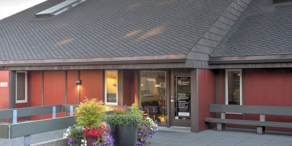
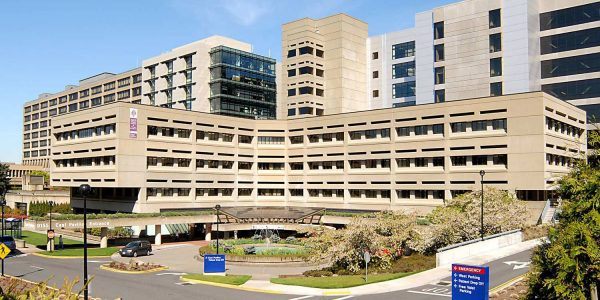
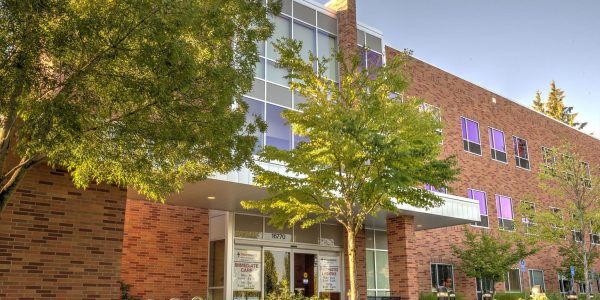
Locations

NW Portland (Suite 210)
-
2230 NW Pettygrove Street, Suite 210
Portland, OR 97210 - (503) 223-6223
- (503) 223-3665

NW Portland (Suite 160)
-
2226 NW Pettygrove, Ste. 160
Portland, OR 97210 - (503) 223-6223
- (503) 546-7228

SW Portland / St. Vincent Hospital
-
9135 SW Barnes Rd #663
Portland, OR 97225 - (503) 297-1078
- (503) 292-2176


